
How should I study Anatomy of Flowering Plants for NEET in six steps?
Study Anatomy of Flowering Plants for NEET by identifying the tissue, locating its arrangement, then testing its distinguishing feature. Build an NCERT comparison sheet for eliminating options. Reconstructing these differences is more useful than repeatedly reading definitions.
- Check the syllabus boundary. Open the current NCERT chapter alongside the official syllabus for your target exam year, available through neet.nta.nic.in. Follow those boundaries rather than assuming every topic in an older textbook or coaching module remains examinable.
- Build a tissue grid. Use four columns: living or dead at maturity, wall characteristics, location, and function. Check these entries against NCERT.
- Parenchyma: living; generally thin cellulose walls; cortex, pith and other ground tissues; storage, photosynthesis when chloroplasts are present.
- Collenchyma: living; uneven corner thickening; beneath the epidermis of young dicot stems; support.
- Sclerenchyma: generally dead at maturity; thick, lignified walls; fibres and sclereids in supporting regions; strength.
- Xylem tracheids and vessels: dead at maturity; lignified walls; xylem; water conduction and support.
- Xylem fibres: dead; thick walls; xylem; support.
- Xylem parenchyma: living; thin cellulose walls; xylem; storage and radial water conduction.
- Phloem sieve-tube elements: living but without a nucleus at maturity; cellulosic walls and sieve plates; angiosperm phloem; food transport.
- Companion cells: living and nucleated; thin walls; beside sieve tubes; maintain sieve-tube function.
- Phloem parenchyma: living; thin cellulose walls; phloem, absent in most monocots; storage.
- Phloem fibres: dead at maturity; thick walls; phloem; support.
Star the exceptions to blanket rules about conducting tissues. Xylem parenchyma is living, phloem fibres are dead, and mature angiosperm sieve-tube elements remain living without a nucleus.
- Attach tissues to systems. The epidermal system includes epidermis, stomata and epidermal appendages. Ground tissues occupy regions such as cortex, pith and mesophyll; the vascular system contains xylem and phloem. Attach each tissue to its location in a section.
- Build one root–stem comparison sheet. Keep these entries together. Use arrangement and xylem orientation before pith size.
- Roots: radial arrangement, with xylem and phloem on different radii; exarch primary xylem, with protoxylem toward the outside.
- Stems: conjoint bundles, with xylem and phloem together; endarch primary xylem, with protoxylem toward the centre.
- Typical dicot stem: ring-arranged, open bundles; cambium is present between xylem and phloem within the bundle.
- Typical monocot stem: scattered, closed bundles; intrabundle cambium is absent.
- Typical dicot root: fewer xylem patches; small or inconspicuous pith.
- Typical monocot root: polyarch xylem, meaning many xylem patches; large pith.
- Compare leaves, then redraw. A typical dorsiventral leaf has palisade and spongy mesophyll, with more stomata below; an isobilateral leaf has undifferentiated mesophyll and stomata on both surfaces. Grass leaves have bulliform cells in the upper epidermis. Keep orientation separate: xylem adaxial, phloem abaxial. Redraw NCERT sections from memory, label diagnostic structures, then check against the book; artistic detail is unnecessary.
- Retrieve before solving. Close NCERT and reconstruct your sheet. Reopen only to correct missing or reversed entries; turn each correction into a self-test question. For every MCQ, mark correct or incorrect, identify the tissue or organ using combined clues, and name the feature that eliminates each distractor.
How do I identify an unfamiliar transverse section?
Use vascular arrangement and xylem orientation before choosing the organ. Then use bundle distribution and cambium to distinguish typical dicot and monocot patterns. A circular outline cannot distinguish roots from stems.
Original practice MCQ, not a previous-year NEET question: A transverse section shows scattered vascular bundles surrounded by sclerenchymatous bundle sheaths. The bundles lack cambium, and the primary xylem is endarch. Identify the section.
- A: Dicot root
- B: Monocot root
- C: Dicot stem
- D: Monocot stem
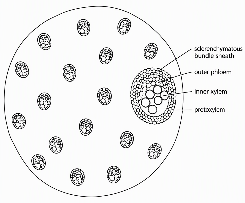
Answer: D, monocot stem. Conjoint bundles with endarch xylem support stem identification. Scattered bundles and absent intrabundle cambium distinguish the typical monocot stem.
Eliminate both roots because their vascular arrangement is radial and their primary xylem is exarch. Eliminate the typical dicot stem because its bundles form a ring and are open.
One vaguely remembered feature is weak evidence; check the combination. Recall task: state the changes for a dicot stem before checking: bundles in a ring, with cambium present within them.
For more section-identification practice, NEET JEEnius AI practice mode offers topic sets that skip questions already seen, with 60 free sets a month.
How do I catch a reversed feature in a leaf MCQ?
Mark “incorrect” before evaluating any statement, then check vascular orientation separately. In the standard NCERT leaf section, adaxial means the upper surface and abaxial means the lower surface. Xylem faces the upper surface; phloem faces the lower one. Recognising three familiar true statements is not enough if you miss “incorrect”.
Original single-correct practice MCQ: Which statement is incorrect about typical NCERT leaf anatomy?
- A: In grass leaves, certain adaxial epidermal cells form bulliform cells.
- B: In an isobilateral leaf, mesophyll is not differentiated into palisade and spongy tissues.
- C: In a leaf vascular bundle, xylem lies toward the abaxial surface and phloem toward the adaxial surface.
- D: A typical dorsiventral leaf has more stomata on its abaxial epidermis than on its adaxial epidermis.
Answer: C. It reverses the tissues. The correct arrangement is xylem adaxial, phloem abaxial; A, B and D are true in their stated contexts.
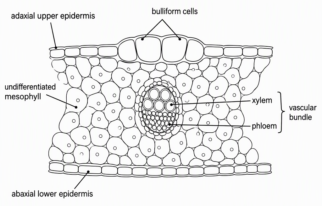
For a quick sketch, mark the upper surface before placing xylem and phloem. Treat bulliform cells as a grass-leaf feature, not a universal leaf feature.
How do I distinguish collenchyma from other supporting tissues?
Combine cell state, wall structure and location, not function alone. Collenchyma and sclerenchyma both provide mechanical support, so “supporting tissue” cannot settle the answer. Living cells with uneven corner thickening point to collenchyma.
Original practice MCQ: A tissue beneath the epidermis of a young dicot stem consists of living cells with uneven thickening at the corners and provides mechanical support. Identify it.
- A: Parenchyma
- B: Collenchyma
- C: Sclerenchyma fibres
- D: Xylem parenchyma
Answer: B, collenchyma. Apply the tissue grid in order. Living cells rule out mature sclerenchyma fibres, but not ordinary parenchyma or xylem parenchyma.
Uneven corner thickening distinguishes collenchyma from both parenchyma options. This thickening involves cellulose, hemicellulose and pectin; sclerenchyma instead has thick, lignified walls. The position beneath a young dicot stem’s epidermis supports the diagnosis.
Xylem parenchyma belongs within xylem and lacks the characteristic corner thickening. Its living state alone does not make it a match.
Change the feature cluster: dead cells with thick, lignified walls would redirect the diagnosis toward sclerenchyma. That alteration turns one solved MCQ into a self-test.
What should I practise after making the anatomy comparison sheet?
Start separately with tissue identification, root–stem comparisons and leaf anatomy. Move to mixed questions once you can reconstruct the sheet without looking.
Practise each format deliberately:
- Unlabelled or partially labelled diagrams: identify the organ and label diagnostic structures.
- Direct feature-identification MCQs: combine cell state, wall type and location.
- Correct/incorrect statement MCQs: mark which is asked, then check each statement.
After these original examples, use genuinely sourced chapterwise NEET previous-year questions. Verify paper attribution and answer keys rather than trusting a “PYQ” label.
Log the mistaken pair, exact NCERT correction and preventive clue:
- Exarch versus endarch: copy the protoxylem-position distinction; outer protoxylem indicates roots, inner indicates stems.
- Open versus closed: copy the cambium distinction; check within the bundle, not between neighbouring bundles.
For a marking scheme illustrated below, three correct answers score:
Two correct answers and one wrong answer score:
One reversal therefore costs five marks compared with answering correctly. A guessed correct answer is not secure until you explain why the other options fail.
At your next session, reproduce the failed diagram or comparison before fresh questions; repeat during later mixed revision. Readiness means labelling standard sections, identifying organs through independent features, and explaining tissue exceptions without notes.
For that next session, NEET JEEnius AI daily practice problems provide a fresh topic set every day, with 20 free attempts a month. Reconstruct the failed comparison before opening the set.
Next step: daily practice problems on NEET JEEnius AI and get a fresh set on a topic every day (20 free attempts a month).
Read next: General Organic Chemistry Practice Questions NEET: 6 MCQs.
Frequently asked questions
How should I study Anatomy of Flowering Plants for NEET?
Check the current NCERT chapter against the official syllabus for your target exam year. Build a tissue grid covering living or dead status, wall characteristics, location and function, then compare roots, stems and leaves. Reconstruct these comparisons and redraw diagnostic sections without notes before solving MCQs.
How do I distinguish a root from a stem in a transverse section?
Use vascular arrangement and primary xylem orientation first: roots have radial arrangement and exarch xylem, while stems have conjoint bundles and endarch xylem. In typical stems, ring-arranged open bundles indicate a dicot, while scattered closed bundles indicate a monocot. A circular outline alone cannot distinguish roots from stems.
Which xylem and phloem cells are living or dead?
Xylem tracheids, vessels and fibres are dead at maturity, while xylem parenchyma is living. Phloem sieve-tube elements, companion cells and parenchyma are living, while phloem fibres are dead at maturity. Mature angiosperm sieve-tube elements remain living despite lacking a nucleus.
Is xylem on the upper or lower side of a leaf vascular bundle?
In the standard NCERT leaf section, xylem faces the adaxial or upper surface, while phloem faces the abaxial or lower surface. Mark the upper surface before placing either tissue in a sketch. In statement MCQs, check this orientation separately and note whether the question asks for the correct or incorrect statement.