Which two structures form JGA in the NEET 2026 question?
Answer D: the distal convoluted tubule and afferent arteriole is correct for the juxta glomerular apparatus NEET 2026 question. Both structures must match the specialised contact site.
The distal segment contacts the vascular region of its own renal corpuscle. This is a local contact within the same nephron, not an opening joining the tubule lumen to the blood vessel.
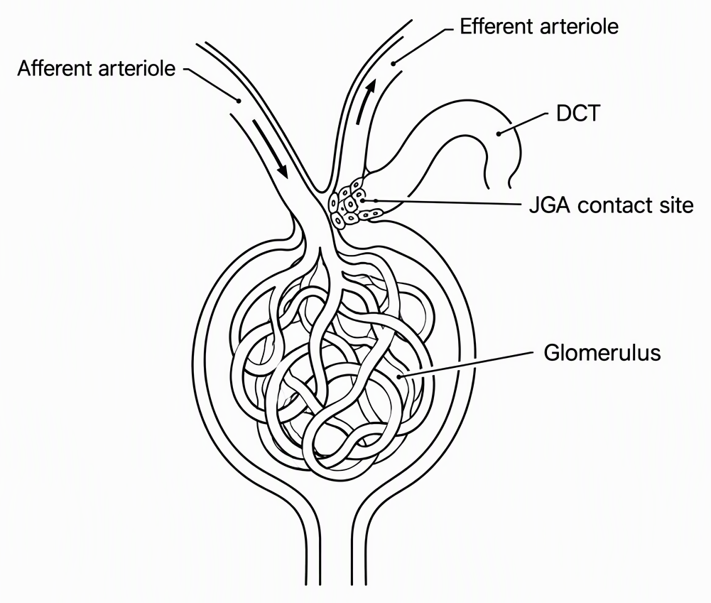
Paraphrased question: In a nephron, specialised cells at the meeting point of which tubular segment and blood vessel form JGA?
Single-correct; question-bank difficulty: medium, tag 2; expected solving time: 30 seconds. The time is a question-bank expectation for this two-part identification, not measured student performance.
How do you solve the JGA question by checking the tubule and arteriole?
Answer D: distal convoluted tubule and afferent arteriole. Check the tubular component first, then its vascular partner. The supplied solution uses contact anatomy: identify the modified cellular regions and match both structures to the choices.
- Identify the tubule. The modified tubular region belongs to the distal convoluted tubule, or DCT. It is not the proximal convoluted tubule, or PCT.
- Remove the PCT options. B and C both name the proximal tubule, so eliminate both. Only A and D remain.
- Identify the blood vessel. The relevant contact is with the afferent arteriole, which carries blood into the glomerulus. The efferent arteriole carries blood away from it.
- Remove the wrong vascular partner. A names the efferent vessel. Eliminate A, leaving D as the only pair that passes both checks.
- Reassemble the explanation. JGA consists of specialised cellular regions of the DCT and afferent arteriole where they meet within the same nephron.
Afferent means entering; efferent means leaving, relative to the glomerulus. That direction cue distinguishes the vessels but does not replace learning the actual contact anatomy. The location and cellular modifications establish the answer; no hormone pathway is needed.
Why is option A wrong even though it contains DCT?
A gets the tubule right but the arteriole wrong. The method error is accepting the whole option after recognising DCT, without independently checking its vascular partner.
The DCT’s later position along the filtrate pathway does not mean it contacts the vessel carrying blood out of the glomerulus. Tubular flow and blood flow are different pathways. “Distal” does not imply “efferent.”
Check DCT or PCT, then afferent or efferent; accept the pair only if both are correct. Recall “DCT meets the entering arteriole”, specifically at the contact within the same nephron.
Under the supplied marking scheme, selecting D earns four marks; selecting A loses one. The score difference is:
This is a marking consequence, not anatomical proof. Use the second check to verify the vessel before selecting the option.
Which specialised cells form the tubular side of JGA?
Macula densa cells form the specialised tubular component. They lie in the distal tubular region at the JGA contact. Juxtaglomerular granular cells instead belong to the afferent arteriolar wall.
Original Human Physiology practice question 1, not a verified PYQ: Which specialised cells belong to the tubular side of JGA?
Answer: A, macula densa cells. Podocytes are associated with the visceral layer of Bowman’s capsule, not the DCT component of JGA. Proximal-tubule brush-border cells name the wrong tubular segment, while granular cells name the vascular side of the apparatus.
What do JG cells release, and how do they respond to a fall in GFR?
JG cells release renin; at the NEET textbook level, a fall in GFR can activate increased renin release. The original question asks where JGA forms. These two original Human Physiology practice questions, not verified PYQs, ask what its cells release and why that matters.
Which substance is released by juxtaglomerular cells?
Renin comes from specialised JG cells in the afferent arteriolar wall. Macula densa cells are the tubular component, not the renin-releasing cells.
Original practice question 2: Which substance do juxtaglomerular cells release?
Answer: A, renin. The releasing cells are the vascular granular cells.
Which response can a fall in GFR initiate through JG cells?
Increased renin release is the correct response. Reduced GFR can activate JG cells; renin initiates the renin–angiotensin mechanism. The resulting response helps raise glomerular blood pressure and restore GFR.
Original practice question 3: At the NEET textbook level, which response can falling GFR initiate through JG cells?
Answer: A. The other choices incorrectly identify the direct JG-cell response.
Practise the two-part check before reading each solution: Free NEET Biology Question Bank: MCQs, PYQs and Mocks.
Frequently asked questions
Which two structures form the juxtaglomerular apparatus?
JGA forms at the contact between specialised cellular regions of the distal convoluted tubule and the afferent arteriole of the same nephron. This is a local contact site, not an opening connecting the tubule lumen to the blood vessel.
Why is DCT and efferent arteriole the wrong answer for JGA?
The pair identifies the correct tubule but the wrong blood vessel. JGA involves the afferent arteriole, which carries blood into the glomerulus, not the efferent arteriole, which carries blood away. Check both components before accepting an option.
Are macula densa cells on the tubular or vascular side of JGA?
Macula densa cells form the specialised tubular component at the JGA contact. Juxtaglomerular granular cells belong to the vascular component in the afferent arteriolar wall.
Which cells release renin in JGA?
Juxtaglomerular granular cells in the afferent arteriolar wall release renin. Macula densa cells are the tubular component, not the renin-releasing cells.
How does JGA respond to a fall in GFR?
At the NEET textbook level, a fall in GFR can activate JG cells to increase renin release. Renin initiates the renin–angiotensin mechanism, whose response helps raise glomerular blood pressure and restore GFR.